 Call us:

MRI Knee Scan or Magnetic Resonance Imaging Knee is a special type of scan that uses magnetic rays to create a detailed image of the knee.
MFine offers you high-quality lab options, and an excellent discount of upto 50%, for your MRI Knee in Bangalore.
MRI Scan Knee in Bangalore by MFine
|
MFine offers a Brain MRI for only ₹ 2, 750, which is significantly lower than the typical market price of over ₹ 7, 500.
Dial ☏08061970525 to avail of this offer
Or click the button below for a callback.
You can book an online doctor consultation after booking the test.
Knee MRI scan costs in Bangalore
Below, we have compiled the discounted rates for the most common knee MRI scans available in Bangalore. Keep in mind that these prices are subject to change; for the latest details, please get in touch with us.
| Center | MRP (in Rs) | MFine price (in Rs) |
| Aarthi Diagnostics, Jaya Nagar (Night Booking) | 2750 | 2750 |
| Aarthi Diagnostics, Indiranagar (Night Booking) | 2750 | 2750 |
| Aarthi Diagnostics, Koramangala | 3650 | 3557.5 |
| Aarthi Diagnostics, Rajaji Nagar | 3650 | 3557.5 |
| Aarthi Diagnostics, Padmanabanagar | 3650 | 3557.5 |
| Aarthi Diagnostics, Indiranagar | 3650 | 3557.5 |
| Aarthi Diagnostics, Jaya Nagar | 3650 | 3557.5 |
| Aarthi Diagnostics, Jaya Nagar (Night Booking) | 3750 | 3750 |
| Aarthi Diagnostics, Indiranagar (Night Booking) | 3750 | 3750 |
| Medicover Hospitals, Jayanagar | 7500 | 4300 |
| Aarthi Diagnostics, Padmanabanagar | 4650 | 4507.5 |
| Aarthi Diagnostics, Jaya Nagar | 4650 | 4507.5 |
| Aarthi Diagnostics, Rajaji Nagar | 4650 | 4507.5 |
| Aarthi Diagnostics, Koramangala | 4650 | 4507.5 |
| Aarthi Diagnostics, Indiranagar | 4650 | 4507.5 |
| Aster RV Hospital, JP Nagar | 8000 | 4800 |
| Focus Diagnostics Centre, Rajajinagar | 6800 | 5440 |
| Aarthi Diagnostics, Jaya Nagar (Night Booking) | 5500 | 5500 |
| Aarthi Diagnostics, Indiranagar (Night Booking) | 5500 | 5500 |
| Focus Diagnostics Centre, Rajajinagar | 6800 | 5780 |
Contact us at ☏08061970525 to book a convenient lab appointment at your preferred time.
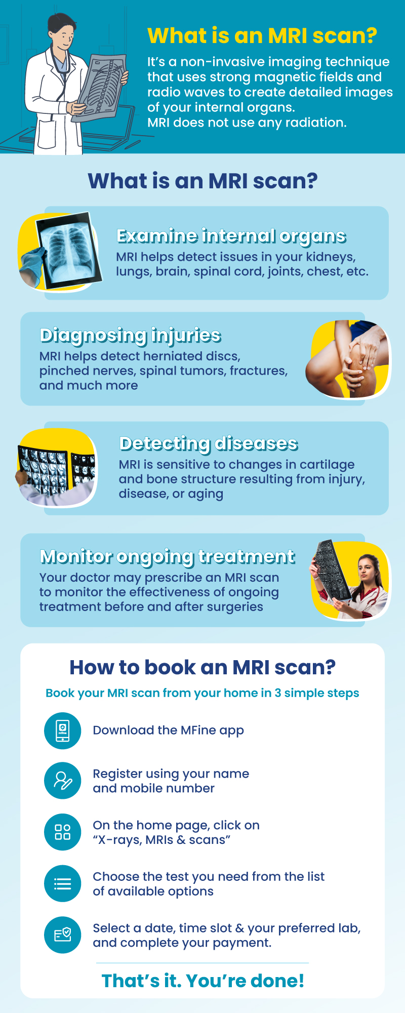
Why should I book MRI through MFine?
|
Exclusive Benefits with MFine
(1) Certified labs
Get access to over 600+ labs certified by NABL and NABH
(2) Same-day slot available
Get scans done on the same day
(3) Quick and convenient
Get reports in 12 hours and digital films in 15 – 20 minutes
(4) FREE Consultation
Post scans, consult a doctor for free to review your report
About Knee MRI Scan
A knee MRI scan helps doctors see images of the knee joint, cartilage, ligaments, muscles, and tendons.
The knee is an intricate joint that links the femur (thigh bone), tibia (shin bone), and patella (knee cap).It is supported by a network of soft tissues, including cartilage, ligaments, tendons, and muscles. A knee MRI scan is a medical imaging method that uses strong magnets and radio waves to create detailed images of the inside of the knee without any invasive procedures. These structures include:
Bones
- Femur (Thigh Bone): The largest bone in the thigh, forming the upper part of the knee joint. It plays a crucial role in supporting body weight and facilitating various knee movements.
- Tibia (Shin Bone): The larger of the two lower leg bones, forming the lower part of the knee joint. It acts as the main weight-bearing bone of the leg.
- Patella (Knee Cap): A small, flat bone located in front of the knee joint, protecting it and providing leverage for knee movement. The patella can be found within the tendon of the quadriceps muscle.
Cartilage
- Meniscus: There are two menisci in the knee – medial and lateral. They are C-shaped cartilage that act as shock absorbers, cushioning the joint during movement. The menisci also help distribute body weight evenly across the knee joint.
- Articular Cartilage: A smooth, protective covering on the ends of bones that allows for smooth joint movement. This cartilage ensures frictionless gliding of the bones during knee motion and protects the joint surfaces from wear and tear.
Ligaments
Ligaments are strong, fibrous bands that connect bones to bones and provide stability to the knee joint. The major ligaments in the knee include:
- ACL (Anterior Cruciate Ligament): The anterior cruciate ligament (ACL) is situated at an angle in the middle of the knee and aids in stabilizing the knee by hindering the tibia from moving excessively forward in relation to the femur.
- PCL (Posterior Cruciate Ligament): The PCL (posterior cruciate ligament) is situated behind the ACL (anterior cruciate ligament) and serves to prevent excessive backward movement of the tibia in relation to the femur.
- MCL (Medial Collateral Ligament): This is located on the inner side of the knee and plays an important role in providing stability to the knee joint and preventing it from bending inward.
- LCL (Lateral Collateral Ligament): This is located on the outer side of the knee and plays a crucial role in providing stability to the joint. It helps to prevent the knee from bending outwards and maintains its proper alignment.
Tendons
Tendons are tough, fibrous tissues that connect muscles to bones. In the knee, they include the:
- Quadriceps Tendon: Connects the quadriceps muscles to the patella. This tendon plays a critical role in knee extension, allowing the leg to straighten.
- Patellar Tendon: Connects the patella to the tibia. It helps transfer the force generated by the quadriceps muscles to the shin bone, enabling movements like jumping and running.
Muscles
Though not technically part of the knee joint, several muscles surround the knee and play a crucial role in knee function and stability. These muscles include:
- Quadriceps: The quadriceps consist of four muscles in the front of the thigh. They work together to extend the knee, allowing the leg to straighten.
- Hamstrings: The hamstrings are three muscles at the back of the thigh. They flex the knee, allowing the leg to bend. They also play a role in hip extension and knee stability.
- Gluteal Muscles (Glutes): The glutes are located in the buttocks and include the gluteus maximus, medius, and minimus. These muscles help position the knee during movement, contributing to overall knee stability.
Joint Capsule
The joint capsule is a thin, tough membrane that surrounds the knee joint, helping to keep it stable. It encloses the joint space and is lined with synovial fluid, which lubricates the joint, reducing friction during movement.
Bursa
Fluid-filled sacs called bursae provide cushioning and reduce friction between tissues in the knee. They help the knee joint move smoothly, preventing irritation and inflammation.
Why Would a Doctor Order a Knee MRI Scan?
A knee MRI scan is recommended for various reasons, including:
Common Injuries:
- Fractures: MRI scans can detect fractures in the bones of the knee joint, helping doctors assess the extent of the injury and plan appropriate treatment.
- Ligament Injuries: Tears or damage to ligaments like the ACL, PCL, MCL, or LCL. MRI scans provide detailed images of ligament injuries, aiding in accurate diagnosis and treatment planning.
- Meniscal Tears: The menisci are susceptible to tears, especially during sports or activities that involve sudden twisting or pivoting movements. MRI scans can identify meniscal tears and guide appropriate treatment strategies.
- Bursitis: Inflammation of the bursae in the knee can cause pain and swelling. An MRI scan helps determine the extent of inflammation and assists in developing a suitable treatment plan.
- Tendonitis: Inflammation of tendons in the knee, such as the patellar tendon or quadriceps tendon, can cause discomfort and limit mobility. MRI scans help visualize tendon inflammation and guide treatment decisions.
- Tendon Tears: Tears or damage to tendons, such as the quadriceps or patellar tendon, can significantly impact knee function. MRI scans provide detailed images of tendon tears, facilitating accurate diagnosis and treatment planning.
- Collateral Ligament Injuries: Injuries to the collateral ligaments on the inner (MCL) or outer (LCL) side of the knee can cause instability. MRI scans help assess the severity of collateral ligament injuries and guide appropriate management.
- Iliotibial Band Syndrome: A common overuse injury that affects the outer part of the knee. MRI scans can help identify inflammation or injury to the iliotibial band, guiding appropriate treatment and rehabilitation.
- Posterior Cruciate Ligament Injuries: Tears or damage to the PCL can occur due to significant trauma or sports-related injuries. MRI scans play a crucial role in diagnosing PCL injuries and planning suitable treatment approaches.
Prerequisites for a Knee MRI Scan
In most cases, there are no specific prerequisites for a knee MRI scan. However, inform your doctor if you have any metal implants or devices in your body, as they may interfere with the MRI procedure. Additionally, let your doctor know about any allergies or medical conditions you may have, as some contrast agents used in MRI scans may not be suitable for certain individuals.
What to Expect During a Knee MRI Scan
If your doctor recommends a knee MRI scan, here’s what you can expect during the procedure:
- Preparation: You will likely be asked to change into a hospital gown to avoid any interference from metal objects in your clothing. You may also need to remove jewelry, watches, and other metallic accessories.
- Positioning: You will lie down on a movable examination table, which will slide into the MRI machine. The MRI technician will ensure that you are comfortably positioned for the scan.
- Stillness: During the scan, it is essential to remain as still as possible to obtain clear and accurate images. The MRI machine will make loud noises during the procedure, but earplugs or headphones may be provided to minimize discomfort.
- Contrast Agent (If Required): In some cases, a contrast agent may be administered intravenously to enhance the visibility of certain structures in the knee. This contrast material helps highlight blood vessels and areas of inflammation.
- Duration: The duration of the MRI scan can vary depending on the complexity of the images required. On average, a knee MRI scan takes between 30 to 60 minutes to complete.
- Comfort: While MRI scans are generally painless, some patients may find the enclosed space of the MRI machine uncomfortable. If you experience claustrophobia or discomfort, inform the MRI technician, who may provide strategies to help you feel more at ease.
FAQs
How long does an MRI last for a knee?
An MRI of the knee typically lasts around 20 to 45 minutes.
Is MRI safe for knee pain?
Yes, MRI is generally safe and commonly used to diagnose knee pain and related issues.
What side effects can you get from an MRI?
MRI usually has no side effects, but some people might experience claustrophobia or mild discomfort from the machine’s noise.
Read more about the side effects of MRI scan: Is MRI safe for everyone?
What will an MRI tell me about my knee?
An MRI can provide detailed images of structures within the knee, helping diagnose injuries, inflammation, and other issues.
Can you MRI both knees at the same time?
Yes, it’s possible to MRI both knees simultaneously if the imaging facility offers appropriate equipment for it.
Other topics you may be interested in:
| For further assistance call us on ☏08061970525 |


 Call
Now
Call
Now