Avail good discounts on your knee MRI scan costs in Mumbai. MFine offers you high-quality lab options and an excellent discount of upto 50% for your MRI Knee in Mumbai.
|
MRI Scan Knee in Mumbai by MFine
|
Looking for a knee MRI in Mumbai?
The usual market rate is above ₹7,000, but we have a special deal for you! For a limited period, you can get your MRI done for only ₹3557.5, saving up to 50%! Don’t hesitate click the button below to request a callback or call us at
Or, you could also request a callback
Get more value for your health with our exclusive deal! When you book your knee MRI in Bangalore, you’ll not only save big on the scan cost (₹3557.5) but also receive a FREE online consultation with a doctor.
Knee MRI scan costs in Mumbai
Below, we’ve gathered the reduced rates for the commonly conducted knee MRI scans in Mumbai. Kindly be aware that these prices could undergo changes; to obtain the latest details, please contact us directly.
| MRI Knee Cost in Mumbai | Offer Price |
| MRI Knee Price in Mumbai | ₹3557.5 |
| MRI Both Knee Price in Mumbai | ₹6740 |
Contact us at ☏08061970525 to book a convenient lab appointment at your preferred time.
Why should I book an MRI through MFine?
|
Exclusive Benefits with MFine
(1) Certified labs
Get access to over 600+ labs certified by NABL and NABH
(2) Same-day slot available
Get scans done on the same day
(3) Quick and convenient
Get reports in 12 hours and digital films in 15 – 20 minutes
(4) FREE Consultation
Post scans, consult a doctor for free to review your report
All about Knee MRI Scan
The knee is a remarkable joint that connects the femur, tibia, and patella, forming a complex network of bones, cartilage, ligaments, tendons, muscles, joint capsule, and bursae. Its intricate structure allows for smooth movement and stability, but it also makes the knee susceptible to various injuries and conditions.

Understanding the Components of the Knee Joint
- Bones:
- Femur (Thigh Bone): As the largest bone in the thigh, the femur plays a crucial role in supporting the body’s weight and facilitating a wide range of knee movements.
- Tibia (Shin Bone): Serving as the larger of the two lower leg bones, the tibia acts as the primary weight-bearing bone of the leg and contributes to knee stability.
- Patella (Knee Cap): Nestled in front of the knee joint, the patella protects the joint and enhances knee movement by functioning within the tendon of the quadriceps muscle.
- Cartilage:
- Meniscus: The knee houses two menisci, the medial and lateral, which are C-shaped cartilage structures. These act as shock absorbers, cushioning the joint during movement and promoting even distribution of body weight.
- Articular Cartilage: A smooth and protective covering on the ends of bones that facilitates frictionless gliding during knee motion and safeguards joint surfaces from wear and tear.
- Ligaments:
- ACL (Anterior Cruciate Ligament): Positioned at an angle in the middle of the knee, the ACL is instrumental in stabilizing the knee by preventing excessive forward movement of the tibia relative to the femur.
- PCL (Posterior Cruciate Ligament): Located behind the ACL, the PCL serves to prevent excessive backward movement of the tibia relative to the femur.
- MCL (Medial Collateral Ligament): Found on the inner side of the knee, the MCL contributes to knee stability and prevents inward bending of the joint.
- LCL (Lateral Collateral Ligament): Situated on the outer side of the knee, the LCL is essential for maintaining proper alignment and preventing outward bending of the knee.
- Tendons:
- Quadriceps Tendon: Connecting the quadriceps muscles to the patella, this tendon is pivotal for knee extension, enabling the leg to straighten.
- Patellar Tendon: Joining the patella to the tibia, this tendon facilitates dynamic movements like jumping and running by transferring the force generated by the quadriceps muscles.
- Muscles:
- Quadriceps: Comprising four muscles at the front of the thigh, the quadriceps synergize to extend the knee and straighten the leg.
- Hamstrings: Positioned at the back of the thigh, the hamstrings flex the knee, allowing the leg to bend. They also contribute to hip extension and knee stability.
- Gluteal Muscles (Glutes): Located in the buttocks region, the gluteal muscles, including the gluteus maximus, medius, and minimus, assist in positioning the knee during movement and promoting overall knee stability.
- Joint Capsule: The knee joint is encapsulated by a thin yet robust membrane known as the joint capsule, which plays a critical role in maintaining joint stability. This capsule is lined with synovial fluid, which lubricates the joint, reducing friction during movement.
- Bursa: The knee joint features fluid-filled sacs called bursae, which provide cushioning and minimize friction between tissues during knee movements. Bursae contribute to the smooth functioning of the knee joint and help prevent irritation and inflammation.
The Significance of a Knee MRI Scan
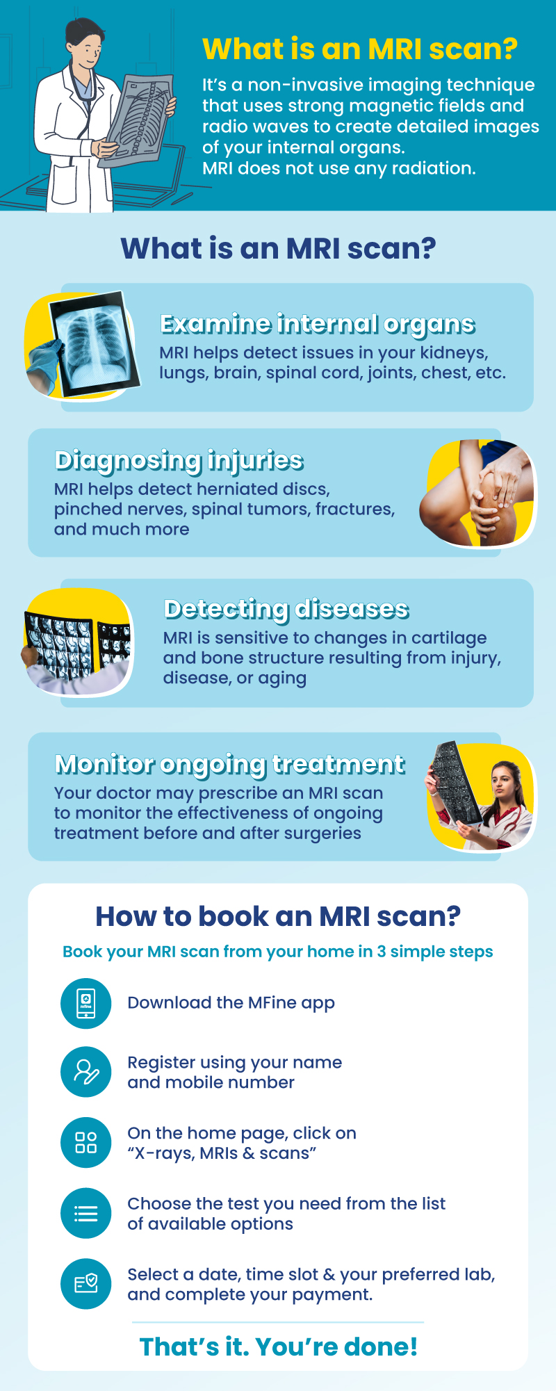
A knee MRI scan is a valuable diagnostic tool used by healthcare professionals for a wide range of knee-related issues. This non-invasive medical imaging procedure utilizes powerful magnets and radio waves to generate detailed, cross-sectional images of the knee’s internal structures. The significance of a knee MRI scan lies in its ability to:
- Accurately Diagnose Injuries: A knee MRI scan can identify and assess various knee injuries, such as fractures, ligament tears (ACL, PCL, MCL, LCL), meniscal tears, bursitis, tendonitis, and collateral ligament injuries. The detailed images obtained from the scan aid in precise diagnosis and appropriate treatment planning.
- Evaluate Degenerative Conditions: Knee MRI scans are instrumental in evaluating degenerative conditions that affect the knee joint, such as osteoarthritis. By visualizing changes in the cartilage and joint space, healthcare professionals can tailor treatment strategies to manage and alleviate symptoms.
- Assess Joint Health: The scan can reveal the overall health of the knee joint, including the condition of the articular cartilage, which is essential for ensuring smooth joint movement and preventing degeneration.
- Plan Surgical Interventions: In cases where surgical intervention is necessary, a knee MRI scan provides surgeons with critical information about the extent of the injury or condition, enabling them to plan and execute procedures with precision.
Reasons for Ordering a Knee MRI Scan
A knee MRI scan is recommended for various reasons, including:
- Traumatic Injuries: MRI scans are valuable in diagnosing and assessing the extent of traumatic injuries to the knee joint, such as fractures, ligament tears, and meniscal tears.
- Chronic Knee Pain: Patients experiencing chronic knee pain, swelling, or limited mobility may undergo a knee MRI scan to identify the underlying cause of their symptoms.
- Sports Injuries: Athletes or individuals participating in sports activities that involve repetitive knee movements and impact are prone to knee injuries. A knee MRI scan helps detect these injuries and guides appropriate treatment.
- Degenerative Conditions: Patients with degenerative conditions like osteoarthritis may undergo knee MRI scans to evaluate the severity of joint damage and monitor disease progression.
- Preoperative Evaluation: Before performing knee surgeries, such as ACL reconstruction or meniscal repair, a knee MRI scan is often conducted to assess the condition of the knee joint and aid in surgical planning.
What to Expect During a Knee MRI Scan?
A knee MRI scan is a painless procedure that typically takes between 30 to 60 minutes to complete. Patients can expect the following steps during the scan:
- Preparation: Before the scan, patients may be asked to change into a hospital gown and remove any metallic items such as jewelry, watches, and accessories.
- Positioning: Once prepared, the patient lies down on a movable examination table, which will be slowly guided into the MRI machine.
- Stillness: During the scan, it is essential for patients to remain as still as possible to ensure clear and accurate images. The MRI machine will emit loud noises during the procedure, but earplugs or headphones may be provided to minimize discomfort.
- Contrast Agent (If Required): In some cases, a contrast agent may be administered intravenously to enhance the visibility of certain knee structures. The contrast material highlights blood vessels and areas of inflammation, aiding in a more detailed examination.
- Comfort: While MRI scans are generally well-tolerated, some patients may experience discomfort due to the enclosed space of the MRI machine. If a patient experiences claustrophobia or discomfort, the MRI technician can provide strategies to help them feel more at ease.
FAQs
Where is the normal patella on MRI?
The patella, or kneecap, is located in the front of the knee joint and can be clearly seen on an MRI of the knee.
Is MRI necessary for knee pain?
An MRI can be valuable for diagnosing the cause of knee pain when other methods are inconclusive, helping identify underlying issues in detail.
Is a knee MRI full body?
No, a knee MRI specifically focuses on the knee joint and surrounding structures. It doesn’t encompass the entire body.
Can I eat before a knee MRI?
Yes, you can usually eat before a knee MRI. However, your healthcare provider might recommend avoiding heavy meals if you’ll be receiving contrast dye.
How long do knee MRI results take?
MRI results for the knee can take anything between 15 to 30 minutes.
Read more on the side effects of an MRI scan.
Other topics you may be interested in
| For further assistance call us on ☏08061970525 |

 Call us:


 Call
Now