 Call us:

Limited-time discount: Save big on Spine MRI scans in Kolkata while still being able to access high-quality lab services.
MFine offers you high-quality lab options, and an excellent discount of upto 50%, for your MRI Spine in Kolkata.
MRI Scan Spine in Kolkata by MFine
|
Generally, the market price of an MRI spine scan is above ₹8000 but with us, you can get it for ₹5100 only.
Avail of this exclusive offer by calling us on
Or you can click on the button below for us to call you back.
You can book an online doctor consultation after booking the test.
Spine MRI scan costs in Kolkata
Stay informed about the commonly available Brain MRI scans in Kolkata and their discounted rates as mentioned below. Please be aware that prices are subject to change; feel free to reach out to us for the most current pricing.
MRI Scan Spine Cost in Kolkata |
Offer Price |
| Whole MRI scan spine cost in Kolkata |
₹13500 |
| Cervical MRI scan spine cost in Kolkata | ₹5400 |
| MRI Dorsal Spine Price in Kolkata | ₹5400 |
| MRI Lumbar Spine Price in Kolkata | ₹5400 |
Dial ☏08061970525 to talk to us.
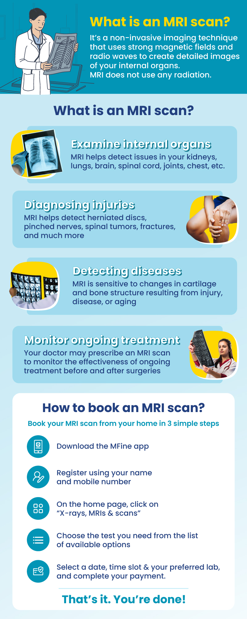
Why should I book MRI through MFine?
|
Exclusive Benefits with MFine
(1) Certified labs
Get access to over 600+ labs certified by NABL and NABH
(2) Same-day slot available
Get scans done on the same day
(3) Quick and convenient
Get reports in 12 hours and digital films in 15 – 20 minutes
(4) FREE Consultation
Post scans, consult a doctor for free to review your report
MRI of the Spine
MRI of the spine captures detailed images of the spine and its neighboring tissues. Let’s now examine the individual parts and segments of the spine.
- Vertebrae: The spine consists of 33 vertebrae, divided into five regions: cervical, thoracic, lumbar, sacral, and coccygeal. Each vertebra has a unique structure and function, and they are stacked on top of one another to create the spine’s distinctive curvature. The vertebrae provide structural support to the body and play a crucial role in maintaining posture and bearing the body’s weight.
- Facet Joints: Positioned between adjacent vertebrae, facet joints are synovial joints that enable spinal flexibility and movement. These joints are coated with cartilage, which reduces friction during articulation, allowing for smooth, pain-free movements such as bending, twisting, and stretching.
- Intervertebral Disks: Found between each pair of vertebrae, intervertebral disks are flat, round cushions composed of a tough outer layer called the annulus fibrosus and a gel-like inner core known as the nucleus pulposus. These disks act as shock absorbers, absorbing the impact of everyday activities like walking, running, and jumping. The nucleus pulposus provides elasticity, while the annulus fibrosus offers stability to the disk.
- Spinal Cord and Nerves: The spinal cord, a bundle of nerves, runs through the vertebral column’s central canal. It serves as a vital communication pathway, relaying sensory information from the body to the brain and transmitting motor signals from the brain to muscles and organs. The spinal cord’s protection within the bony vertebral column is paramount, as any damage to it can lead to severe neurological complications.
- Soft Tissues: Complementing the bony and neural structures of the spine, soft tissues play a crucial role in maintaining spinal alignment, providing support, and enabling bodily movements.
- Ligaments: These tough, fibrous bands connect bone to bone, providing stability to the spine and preventing excessive movement. Ligaments play a crucial role in maintaining proper spinal alignment and preventing injuries.
- Muscles: The muscles surrounding the spine play a pivotal role in supporting the body’s weight, controlling posture, and facilitating movements. Strong and flexible back and core muscles are essential for spinal health and overall functional mobility.
- Tendons: Tendons are tough connective tissues that attach muscles to bones. They transmit the forces generated by muscles during movement to the bones, allowing for coordinated and controlled movements.
What are the different spine segments?
The spine, also known as the vertebral column, is the long, flexible structure that runs down the center of your back. It plays a crucial role in supporting your body, protecting the spinal cord, and allowing you to move in various directions. The spine is made up of several sections and bones, each with its specific functions.
(1) Cervical (neck):
- Includes 7 vertebrae labeled as C1 to C7.
- Located in the neck area.
- Enables movements of the neck, allowing you to nod, turn your head, and look around.
- Guards the neural pathways that connect the brain to the rest of the body.
(2) Thoracic (middle back):
- Consists of 12 thoracic vertebrae, named T1 to T12.
- Found in the middle back region.
- Provides support for the middle back and chest area, protecting vital organs like the heart and lungs.
- Helps maintain proper posture and stability.
(3) Lumbar (lower back):
- Comprises 5 vertebrae, labeled L1 to L5.
- Located in the lower back region.
- Offers stability and strength to support your body weight.
- Allows you to bend, twist, and perform various movements.
(4) Sacrum:
- A triangular bone formed by the fusion of 5 sacral vertebrae.
- Links the spine to the hip bones (pelvis).
- Provides stability to the pelvic region, helping to transfer weight between the upper body and legs.
(5) Coccyx (tailbone):
- Composed of coccygeal vertebrae situated at the end of the spine.
- Serves as an anchor for various muscles and ligaments in the pelvic area.
- Although small, it still plays a role in supporting your body while sitting.
How the Spine Works Together:
The spine works as a flexible and interconnected system. The vertebral column has small spaces between each vertebra, allowing it to move and bend. This flexibility permits you to perform various activities, such as walking, running, bending, and lifting objects.
Moreover, the spine’s design protects the spinal cord, which is a bundle of nerves responsible for transmitting messages between your brain and the rest of your body. The spinal cord runs through a hollow canal in the center of the vertebrae, safeguarding it from potential damage.
Taking care of your spine is essential for overall health and well-being. Proper posture, regular exercise, and avoiding excessive strain on your back can help keep your spine in good shape and prevent potential issues in the future. If you ever experience persistent back pain or discomfort, it’s essential to consult a healthcare professional for proper evaluation and treatment.
Which disorders can be diagnosed using a spine MRI?
Utilizing advanced imaging technology, a spine MRI can effectively diagnose a broad spectrum of conditions, such as:
- Arthritis, notably ankylosing spondylitis impacting spinal joints
- Back strains and sprains resulting from injuries or overuse
- Birth defects like spina bifida affecting spinal development
- Bone spurs, abnormal bony growths affecting spinal structures
- Curvatures of the spine, including scoliosis and kyphosis
- Neuromuscular diseases like ALS affecting nerve and muscle function
- Nerve injuries such as spinal stenosis, sciatica, and pinched nerves
- Osteoporosis leading to weakened and fragile bones
- Spinal cord injuries involving fractures, herniated disks, and paralysis
- Spine tumors and cancerous growths within the spinal region
- Spine infections such as meningitis and osteomyelitis
Why would a doctor prescribe a spine MRI?
Prescribing a spine MRI, doctors aim to:
- Diagnose and evaluate spinal conditions, including arthritis, bone spurs, and curvatures (scoliosis, kyphosis).
- Assess and monitor back injuries such as strains, sprains, and fractures.
- Investigate nerve-related problems like spinal stenosis, sciatica, and pinched nerves.
- Detect and evaluate spinal cord injuries, herniated disks, and spinal tumors.
- Diagnose infections like meningitis and osteomyelitis that impact the spine.
Prerequisites for a Spine MRI
To undergo a spine MRI, it’s essential to meet certain prerequisites, including:
- Medical referral: Patients typically require a doctor’s prescription or referral to schedule a spine MRI.
- Pre-scan questionnaire: Completing a questionnaire helps ensure the MRI’s safety and appropriateness for the patient.
- Medical history disclosure: Informing the healthcare provider about any pre-existing medical conditions, allergies, or previous surgeries is crucial.
- Metal object removal: Patients should remove metal objects like jewelry, watches, piercings, and hearing aids to avoid MRI interference.
- Pregnancy status: Female patients must notify the healthcare provider if they are pregnant or suspect pregnancy, as MRI scans are generally avoided during pregnancy, especially in the first trimester.
FAQs
Can MRI see spinal cord damage?
Yes, MRI can visualize and detect spinal cord damage. It can reveal abnormalities such as compression, inflammation, tumors, or other injuries affecting the spinal cord. MRI is an essential tool for evaluating conditions like spinal cord injury, multiple sclerosis, and other spinal cord-related disorders.
Can a damaged spinal cord heal?
The ability of a damaged spinal cord to heal depends on the extent and severity of the injury. In cases of mild or partial injuries, some recovery may be possible with rehabilitation and therapy. However, severe spinal cord injuries often result in permanent damage and may lead to varying degrees of permanent disability.
How long does it take to fully recover from a spinal cord injury?
The recovery timeline for a spinal cord injury varies widely depending on the severity of the injury and the individual’s response to treatment and rehabilitation. Some people may experience improvements in function over several months or years, while others may have long-term or permanent disabilities.
Can a lumbar spine MRI show cancer?
Yes, a lumbar spine MRI can show signs of cancerous growths or tumors in the lumbar region. The MRI can reveal abnormal masses or lesions in the spine, which may be further evaluated for malignancy with additional tests, such as biopsy.
Does a lumbar spine MRI show the pelvis?
A lumbar spine MRI primarily focuses on the lumbar vertebrae and surrounding structures, so it may capture a portion of the pelvic region. However, for a comprehensive evaluation of the pelvis, specific imaging techniques like pelvic MRI or CT scans may be necessary.
Read more about MRI scan side effects.
Other topics you may be interested in
| For further assistance call us on ☏08061970525 |


 Call
Now
Call
Now